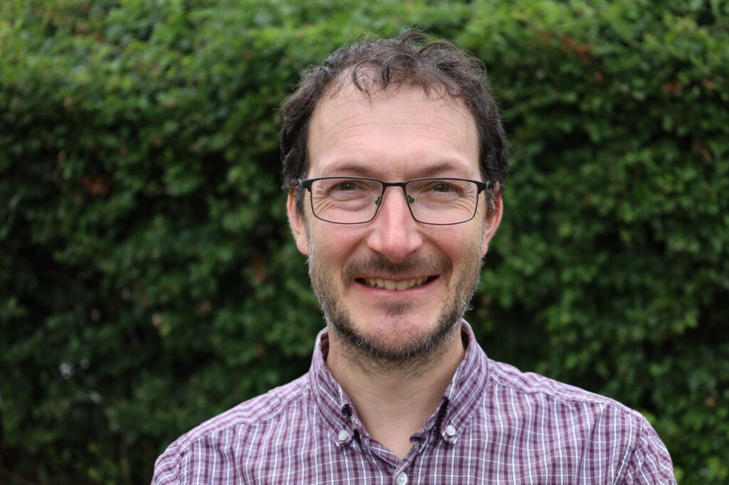
Professor Franck Vidal, from CCPi, has been awarded 3rd place at the Dirk Bartz Prize for Visual Computing in Medicine and Life Science 2025 for a paper which reviewed the use of gVXR in medicine and life sciences.
He shares this award with his co-authors Shaghayegh Afshari, Alberto Albiol, Francisco Albiol, Alberto Corbí, Anna Louise Brun, Chengy-Ying Chou, Pascal Desbarats, Marcos García, Jean-François Giovannelli, Clémentine Hatton, Audrey Henry, Graham Kelly, Claire Michelet, Radu Mihail, Ali Rouwane Hervé Seznec, Aarón Sújar, Jenna Tugwell-Allsup and Pierre-Frédéric Villard.

The winning paper is entitled ‘X-ray simulations with gVirtualXray in medicine and life sciences’ and is available to read here. The paper focuses on VirtualXray (gVXR), a programming interface framework to simulate realistic X-ray projections in realtime on graphics processing units (GPUs). It solves the Beer-Lambert law (attenuation law) using a deterministic X-ray simulation algorithm based on 3D computer graphics, namely rasterisation. Implemented as multi-pass rendering makes it more computationally optimal than the ray-tracing technique, which is a brute-force and straightforward approach to simulate X-ray images.
Although written in C++ using OpenGL and its shading language (GLSL) to leverage the GPU, gVXR is available for other programming languages such as Python. Extensive validation studies, including comparisons with Monte Carlo simulations and real experimental data, have confirmed the accuracy of gVXR’s simulations. gVXR was initially used in medical virtual reality (VR) for training purposes. It was then used in medical physics, and high-throughput data applications including mathematical optimisation and machine learning (ML). Micro-imaging studies on the C. elegans biological model are also reported.
The paper was presented at EuroVis 2025 in Luxembourg. The Dirk Bartz Prize is a biannual competition organised by the Eurographics Association, the Europe-wide professional Computer Graphics association. The award acknowledges the contribution of computer graphics and visualisation techniques; weight is put on demonstrating the impact of the work.
Professor Vidal says:
“It’s been a real pleasure working with users of our software, and see the novel real-world applications it enabled in medicine and life science, from education, to a new type of radiography imaging, improving the diagnosis of lesions in lungs using AI, material decomposition in the emerging spectral computed tomography, to cite a few applications.”
The GVirtualXRay open-source software is available at https://sourceforge.net/projects/gvirtualxray and has been recognized with a Community Choice award by SourceForge.
